Fin regeneration
Fin regeneration
Zebrafish possess the remarkable capability to regrow lost appendages, such as their fins. During this process, different types of cells and tissues, such as bones, skin, nerves and blood vessels reform in a relatively short time frame of about 3 weeks. We are interested in understanding how the growth of new blood vessels occurs during tissue regeneration. We also want to understand if different mechanisms control the blood vessel growth during normal development and tissue regeneration. In order to do so, we use an intubation-based anesthesia protocol that allows us to continuously film regrowing zebrafish fins for up to 2 days. We are also analyzing different zebrafish mutants, in which regenerative blood vessel growth is impaired. We found that regenerating fin arteries are mainly derived from pre-existing veins. Similar to our observations in zebrafish embryos, we also find that the chemokine receptor cxcr4 is instrumental for the correct morphogenesis of these newly forming arteries. Thus, the function of cxcr4 appears to be conserved between embryonic and regenerative blood vessel growth. Currently, we are investigating the role of mural cells during fin blood vessel patterning and establish new artery injury protocols.

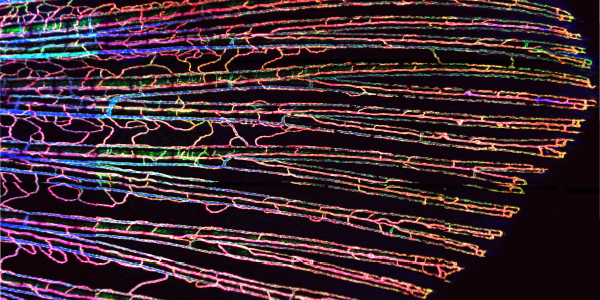
Blood vessels in the fin of an adult zebrafish
We found that mural cells in the zebrafish fin display different morphologies, with mural cells in regions closer to the fish body wrapping around arteries, while those in more distal regions showing long extensions along blood vessels. These morphologies are similar to those of mural cells found in other animals, such as mice. We also found that signaling through platelet derived growth factor beta (pdgfb) was important for the formation of new mural cells. During artery regeneration, new mural cells were not derived from pre-existing ones, implying distinct precursor populations for newly forming mural cells. Together, our results suggest a limited dedifferentiation capacity of established mural cells. In the future we will investigate pathways important for mural cell function and how they might influence the growth of regenerating blood vessels.

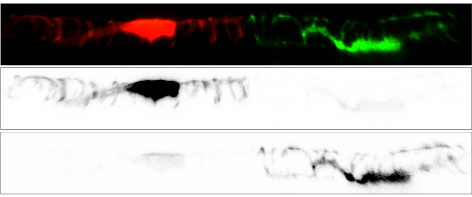
Individually labelled mural cells wrapping around an artery in the zebrafish fin showing their intricate shapes.
Publications
Leonard E. V. , Figueroa R. J., Bussmann J., Lawson N. D. , Amigo J. D. , Siekmann A. F. (2021). Regenerating vascular mural cells in zebrafish fin blood vessels are not derived from pre-existing ones and differentially require pdgfrb signaling for their development. bioRxiv 2021.03.27.437334; doi: https://doi.org/10.1101/2021.03.27.437334
Xu C., Volkery S., Siekmann A.F. (2015). Intubation-based anaesthesia protocol for long-term time-lapse imaging of adult zebrafish. Nature protocols, 10(12):2064-73.
Xu C., Hasan S.S., Schmidt, I., Rocha, S.F., Pitulescu, M.E., Bussmann, J., Meyen, D., Raz, E., Adams, R.H., Siekmann, A.F. (2014). Arteries are formed by vein-derived endothelial tip cells. Nature communications, 5, 5758.

